Fig. 2.
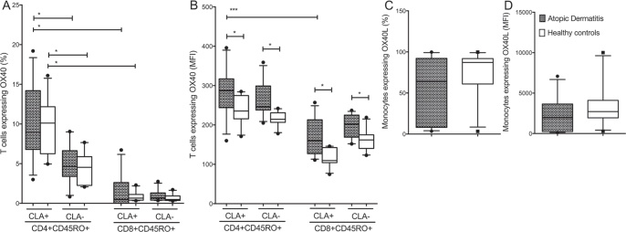
CD4+CD45RO+CLA+ T cells express high levels of OX40 in atopic dermatitis (AD), whereas OX40L is expressed primarily by monocytes. Membrane expression of OX40 and OX40L on peripheral blood mononuclear cells from patients with AD (n = 11) and healthy controls (HC) (n = 10), divided into CD4+CD45RO+ and CD8+CD45RO+ T cells with or without the expression of cutaneous lymphocyte-associated antigen (CLA). (a): OX40 was expressed primarily by the CD4+ CD45RO+ CLA+T cell subsets. (b) Median fluorescence intensity (MFI) of OX40 was significantly higher among patients with AD compared with HC. (c, d) OX40L+ monocytes, showed no difference between patients with AD and HC (both percentage and MFI). Data were analysed as non-paired data by Mann-Whitney U test. Boxes indicate median and interquartile range, and whiskers represent 10th–90th percentiles. *p < 0.05, ***p < 0.001.

