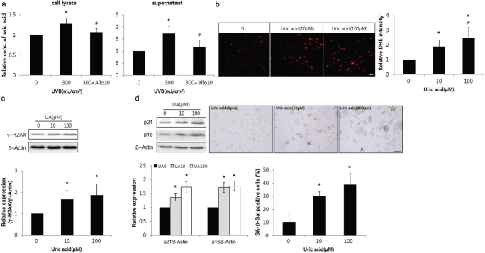
Fig. 5.
Uric acid played the same role as guanine deaminase (GDA) in ultraviolet (UV)-induced keratinocyte (KC) senescence. (a) Assay for uric acid concentration in primary cultures of normal human skin keratinocytes with or without UVB irradiation and allopurinol. (b-d) dihydroethidium (DHE) staining (b) and western blot analysis showing the relative ratios of γ-H2AX (c), p16 and p21 levels and immunofluorescence staining with senescence-associated (SA) β-Gal (d) in KCs with or without exposure to different doses of uric acid. Staining intensities were measured using Wright Cell Imaging Facility (WCIF) ImageJ software. β-Actin was used as an internal control for western blot analysis. Data represent means ± standard deviation (SD) of 3-4 independent experiments. *p < 0.05 vs. control KCs, #p < 0.05 vs. UV-exposed keratinocytes.

