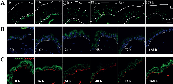
Fig. 1.
Alterations in nerve fibre distribution, and nerve growth factor (NGF) and Sema3A expression in the epidermis of acetone-treated dry skin model mice. (A) Sequential alteration of intraepidermal nerve growth in acetone-treated mice was examined by immunohistochemistry using an anti-PGP9.5 antibody. (B, C) Maximum expression of NGF (green) was noted 16–24 h after the treatment (B). In contrast, the expression level of Sema3A (green) was decreased 24 h after acetone treatment (C). These expression levels gradually returned to normal by 168 h after the treatment. Nuclei are counterstained by DAPI (blue). The broken lines in panel A indicate the border between the epidermis and dermis (basement membrane). The basement membrane in panel B was stained with an anti-nidogen antibody (red). White and broken lines indicate the skin surface and the border epidermis and dermis (basement membrane), respectively. Arrowheads indicate epidermal nerve fibres (green). Scale bars: 15 μm.

