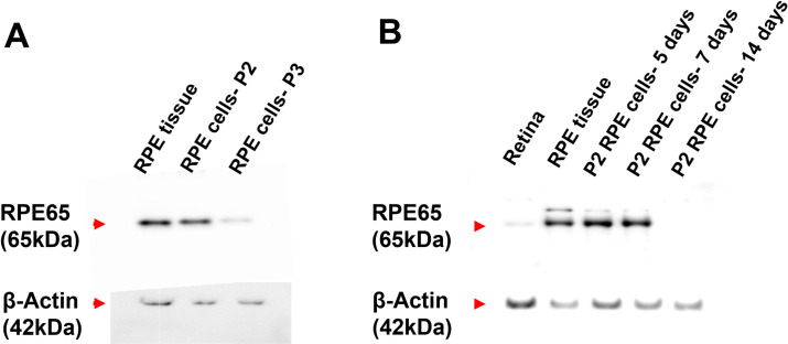
Fig 4. Characterisation of cultured primary porcine RPE cells.
(A) The expression of RPE marker RPE65 in different passages of the cultured primary RPE cells. After 7 days of culture, passage 2 RPE cells showed higher expression of RPE65 than passage 3. RPE tissue was used as a positive control. (B) The expression of RPE65 in passage 2 RPE cells cultured for 5, 7 and 14 days. The passage 2 RPE cells expressed RPE65 up to 7 days, but the expression was absent after 14 days of culture. Whole tissues of RPE and retina were used as positive and negative controls respectively. (n = 3).

