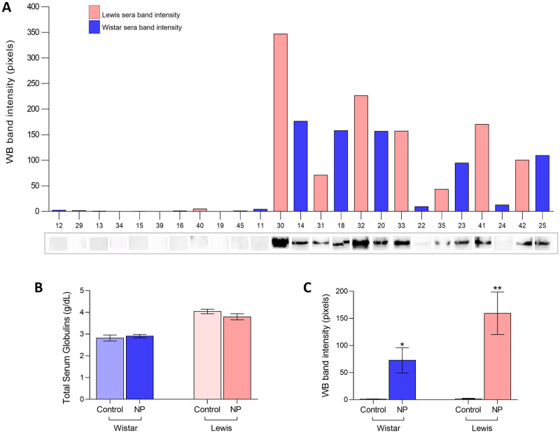
Fig 3.
Immunization: A) Illustrative WB bands, pointing the presence of anti-NP specific IgG antibodies in the serum samples of both Wistar and Lewis rats submitted to NP inoculations (numerical identification of each animal is on X axis), B) Total serum globulin concentration (g/dL), C) WB band intensity quantification.

