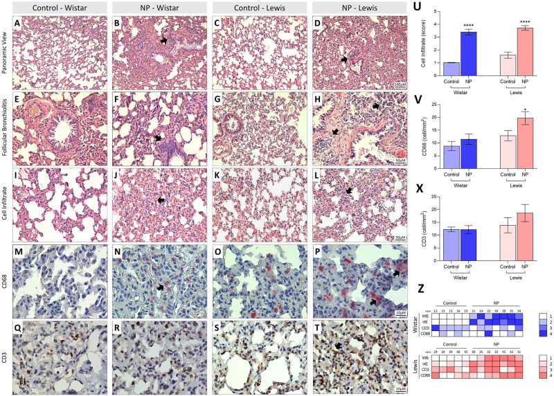
Fig 4.
Histological assessment and immunohistochemistry in lung tissue stained with HE (A-L) and immunostaining by CD68 and CD3 (M-T). In A and C the lung parenchyma presents normal appearance, as well as regular alveolar septa and vessels in the Wistar and Lewis Control group. In contrast, in B and D we identified distortion of pulmonary histoarchitecture and intense thickening in the alveolar septa region with the presence of cellular infiltrate and collagen deposition around the septa in the NP Wistar and Lewis groups (Arrows). Note in F and H the follicular bronchiolitis with the presence of intense lymphocytic infiltrate in the NP Wistar and Lewis groups when compared to the respective controls (E and G). Observe in J and L the thickening of pulmonary interstitium of the NP Wistar and Lewis groups (arrows) when compared to the controls group (I and K). In N we identified in pinkish red the immunostaining for CD68 in the pulmonary interstitium of the NP Wistar group, showing a slight number of cells positive for CD68 and when compared with its respective control (M). In contrast, we observed an increase in cells immunostained for CD68 in lung parenchyma of NP Lewis group (P) when compared to its control group (O). In R, note in brown the immunostaining for CD3 lymphocyte in the lung parenchyma NP Wistar group, when compared to the control group (Q) and in T, we identified CD68-positive cells in the thickening of pulmonary interstitium and control (S). In (U), graphical representation of the score for inflammatory cells indicate significant increase in NP Lewis and Wistar groups. In (V) CD68 statistical difference in Lewis NP and its control significant expression in alveolar septa and peribronchial region and (X) Graphical representation of CD3 quantification of CD3 in alveolar septa and peribronchial region of lung tissue. In Z, note intensity correlation of the anti-NP antibodies and expression of CD68 and CD3 in lung tissue of NP Wistar and Lewis group.

