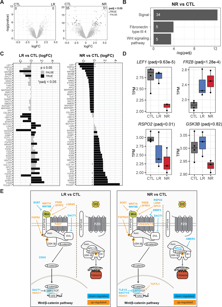
Fig. 2.
Dysregulation of Wnt/β-catenin signaling in NR neurons. a Differential expression results from sorted CTL and LR neurons (left) or sorted CTL and NR neurons (right). Numbers denote DEGs after padj < 0.05. b Top functional terms with Benjamini p < 0.05 from genes identified from NR vs CTL. Numbers indicate the gene count of differentially expressed genes in the category. c Barplots of logFC between LR and CTL or NR and CTL for all Wnt genes (canonical-GO:0060070, and non-canonical-GO:0035567) that were significant with a p raw < 0.05 from at least one comparison. * = padj < 0.05. d Boxplots for key Wnt-signaling related genes. e Graphical representation of the Wnt/β-catenin pathway and the genes showed in c.

