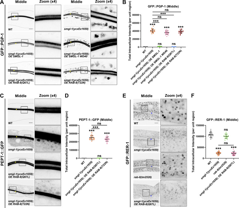
Figure 5.
Expression of constitutively active RAB-8 alleviates the secretion defects of PGP-1 and PEPT-1 caused by SMGL-1 deficiency. (A and B) Confocal images of intestinal cells expressing GFP-tagged PGP-1. Compared with wild-type animals, overexpression of the WD40 domain of SMGL-1 failed to rescue the intracellular accumulation phenotypes of GFP::PGP-1 in smgl-1(ycxEx1659) mutants. Overexpression of RAB-8(Q67L), but not RAB-8(T22N), restored the intracellular accumulation phenotypes of GFP::PGP-1 in smgl-1(ycxEx1659) mutants. (C and D) Confocal images of intestinal cells expressing GFP-tagged PEPT-1. Compared with wild-type animals, overexpression of RAB-8(Q67L), but not RAB-8(T22N), restored the intracellular accumulation phenotypes of PEPT-1::GFP in smgl-1(ycxEx1659) mutants. (E and F) Confocal images of intestinal cells expressing GFP::RER-1. GFP::RER-1 normally resides in the intracellular punctate structures. In smgl-1(ycxEx1659) mutants, GFP::RER-1 appeared diffusive and labeled small puncta. Loss of RAB-8 did not affect the localization of GFP::RER-1, and overexpression of RAB-8(Q67L) failed to restore the localization of GFP::RER-1 in smgl-1 mutants. The signals from the apical membrane were avoided by manual ROI selection. Data are shown as mean ± SD (n = 18 each, six animals of each genotype sampled in three different unit regions of each intestine defined by a 100 × 100 [pixel2] box positioned at random). For multiple comparisons, statistical significance was determined using a one-way ANOVA followed by a post-hoc test (Dunn’s Multiple Comparison Test). ***, P < 0.001. Data distribution was assumed to be normal but this was not formally tested. Scale bars: 10 μm. Colored asterisks indicate intestinal lumen.

