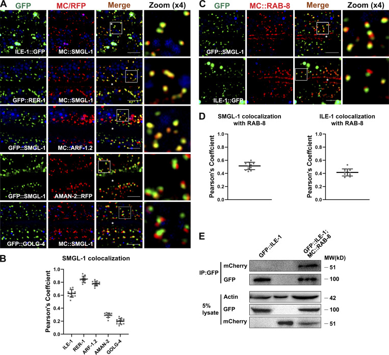
Figure 6.
SMGL-1 resides in the ER-Golgi intermediate compartment and adjacent RAB-8-positive structures. (A and B) Confocal images showing colocalization between SMGL-1 and organelle markers in the intestinal cells. SMGL-1 overlapped with the ERGIC marker ILE-1/ERGIC-53 and the cis-Golgi-located retrograde cargo RER-1 and ARF-1.2/Arf1. SMGL-1 appeared closer in proximity to AMAN-2-labeled cis-/medial-Golgi than to GOLG-4-labeled TGN. (C and D) Confocal images showing colocalization between RAB-8 and SMGL-1 or ERGIC marker ILE-1/ERGIC-53 in the intestinal cells. SMGL-1 often overlapped with RAB-8 in punctate structures. Also, RAB-8 partially colocalized with ILE-1. Pearson’s correlation coefficients for GFP and mCherry signals were calculated (n = 12 animals). The signals from the apical membrane were avoided by manual ROI selection. Scale bars: 10 μm. White asterisks indicate intestinal lumen. (E) In a co-immunoprecipitation assay, ILE-1 precipitated with wild-type RAB-8. Source data are available for this figure: SourceData F6.

