Figure S1.
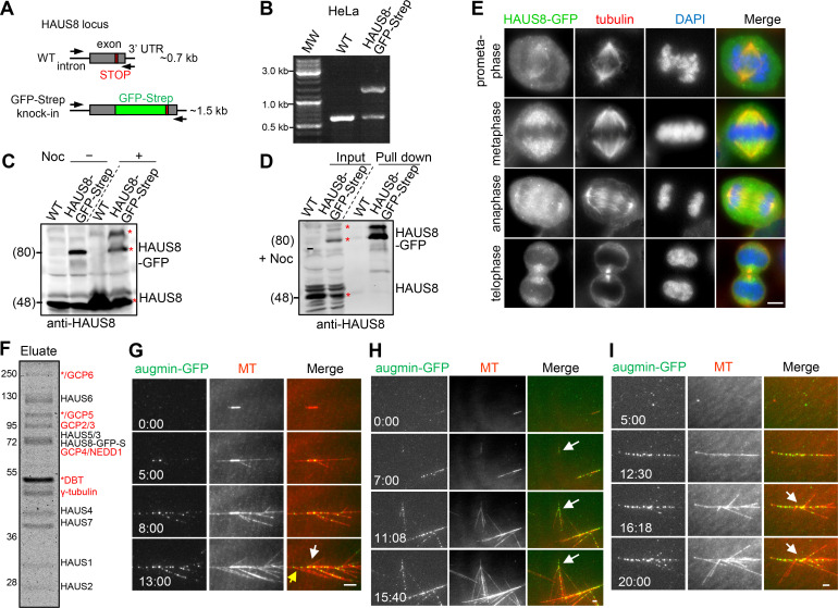
Characterization of HAUS8-GFP-Strep knock-in HeLa cell lines. (A–C) Characterization of HAUS8 knock-in HeLa cell line. (A) Schematic illustration of primer sets and the expected PCR products. PCR genotyping (B) and Western blotting analysis (C) showing that the HAUS8-GFP-Strep knock-in HeLa cell line used in this study is heterozygous. The bands corresponding to up-shifted tagged HAUS8 (nocodazole treated group), tagged, and untagged HAUS8 are marked with red asterisks in C. Of note, GFP-tagging may affect the mobility of HAUS8 on the protein gel, as band shift was only apparent for tagged but not untagged HAUS8. (D) StrepTactin pull-down assay with extracts of nocodazole-arrested mitotic control HeLa (WT) and HAUS8-GFP-Strep knock-in HeLa cells, analyzed by Western blotting with HAUS8 antibody. The bands corresponding to untagged HAUS8 (input), tagged HAUS8, and its up-shifted version (pull-down) are marked with red asterisks. (E) Immunofluorescence staining for α-tubulin and DNA (DAPI) in HAUS8-GFP-Strep knock-in HeLa cells during mitosis. (F) Coomassie blue–stained gel with native augmin–γ-TuRC complex purified from nocodazole-arrested mitotic HAUS8-GFP-Strep knock-in HeLa cells using one-step affinity purification with StrepTactin beads. S, Strep-tag. Asterisks indicate putative contaminants. Above the band of γ-tubulin, a strong band marked with an asterisk represents dihydrolipoamide branched chain transacylase (DBT), as determined by mass spectrometry. (G) Representative images of branching MT nucleation both from GMPCPP seed (denoted by white arrow) and from GDP-MT grown from the minus end of seed (denoted by yellow arrow). (H) Representative images of de novo nucleation of branched MTs from solution (white arrows). (I) Representative images of branch formation at obtuse angles (white arrows). Scale bars, 2 μm.

