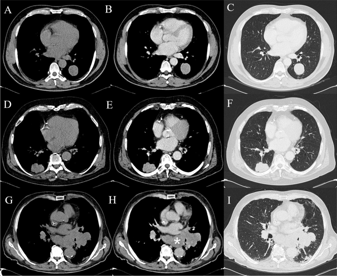
Fig. 2.
Computed tomography imaging in a 67-year-old male patient with a smooth-edged pulmonary lesion with histology of a typical carcinoid in the left lower lobe at baseline (A), with vivid enhancement in the venous phase (B) and lung parenchyma window (C). Large cell carcinoma in a 79-year-old male patient closely connected to the right posterior costal pleura; it shows polylobulated edges at baseline and lung parenchyma window (D, F) with contrast enhancement (E). Small cell carcinoma in a 76-year-old female patient: large left hilar lesion on direct examination (F), with inhomogeneous enhancement (G) and in parenchyma window (H). Notice the multiple confluent mediastinal lymphadenopathies (asterisk)

