Figure 4.
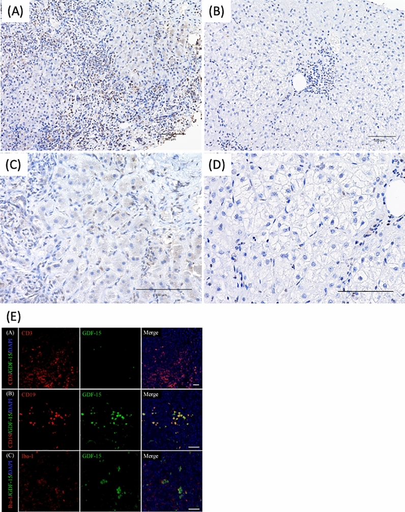
Evaluation of GDF15 immunostaining in liver tissues of AIH patients before (A,C) and after (B,D) treatment (A,C) GDF15 immunostaining revealed hepatic cytoplasm, sinusoidal endothelial cells, and inflammatory cells infiltrating the portal vein area were strongly stained. The immunohistochemistry total score is equivalent to 7. (B,D) GDF15 staining of liver tissues showed fewer positive cells and weaker staining in inflammatory cells infiltrating the portal vein area after treatment than before treatment. The immunohistochemistry total score is equivalent to 2. (E) Double immunofluorescence staining for GDF15 and CD3, CD19, or Iba-1 in the portal area of liver tissues in AIH patient Upper row, immunofluorescence images of GDF15 (green) and CD3 (red). Middle row, immunofluorescence images of GDF15 (green) and CD19 (red). Lower row, immunofluorescence images of GDF15 (green) and Iba-1 (red). The merged images show that GDF15-positive cells are also CD19-positive cells. GDF15, growth differentiation factor 15; AIH, autoimmune hepatitis.

