
Fig. 2.
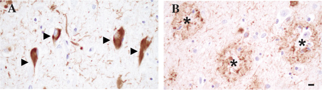
Immunohistochemical stain with anti-phospho tau antibody 7F2 highlighting NFT in the CA1 region of the hippocampal pyramidal cell layer (arrowheads, A) and neuritic plaques in the inferior temporal cortex (asterisk, B). Scale bar = 10 μm, shown in (B)