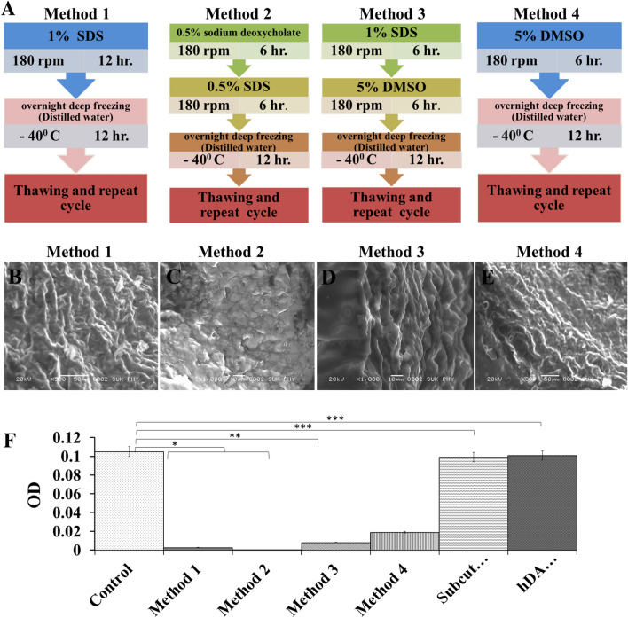
Fig. 1.
A Four different methods used with different chemicals concentration for decellularization of native goat artery. B–E FE-SEM images of decellularized artery (DA) scaffolds obtained after completion of four different decellularization processes. F DNA content in control (Native artery), different scaffolds obtained by different decellularization methods, subcutaneously transplanted DA for 14 days and after transplantation hDA studies. Methods 2 showed complete removal of DNA from scaffold. In vivo experiment showed equal amount of cellular DNA in DA and hDA grafts after transplantation compared to native vessel DNA

