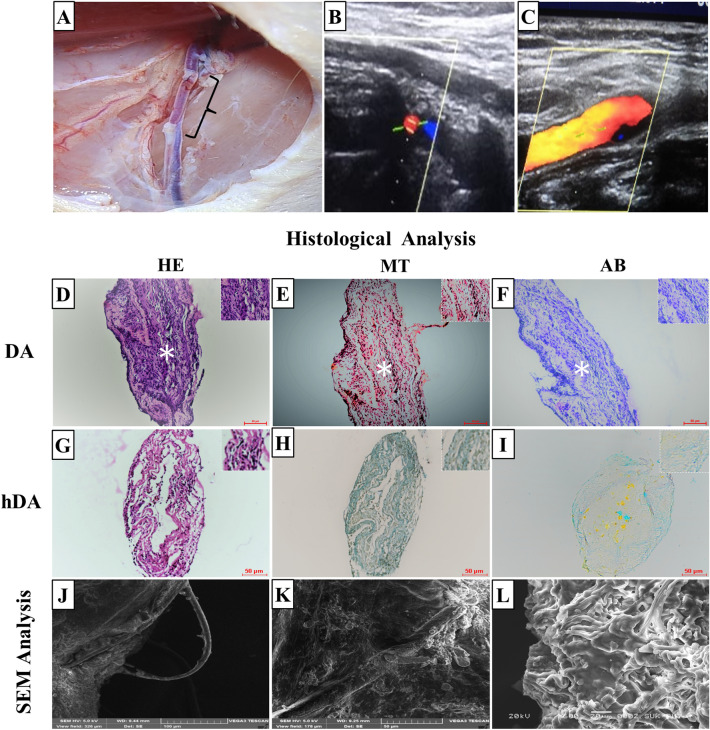
Fig. 3.
A hDA graft transplant in right femoral artery of rat (B, C) color doppler images after one month of transplantation. D–I Histological analysis of DA and hDA transplanted grafts. Hematoxylin Eosin stain (D, G), Masson’s trichrome stain (E, H), and Alcian blue stain (F, I) images of transplanted DA and hDA graft in rat femoral artery. D–F Graft completely occluded with thrombus observed in lumen of DA graft. Thrombus indicated with white artistic. J–L SEM analysis showed cellular lining on surface of transplanted hDA graft

