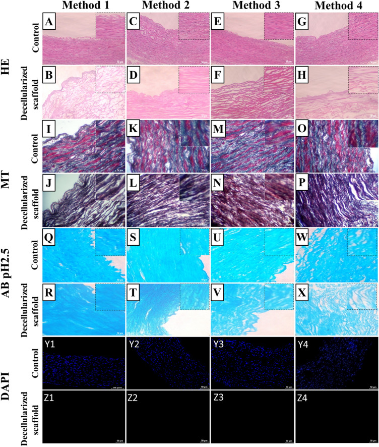
Fig. 4.
Histochemical study of control (native goat artery) and decellularized scaffolds obtained by different methods. (A to H) HE image of native tissue and complete cell free DA scaffolds. In method 2 decellularized artery scaffold (D) showed intact intimal and medial structure compared to other three methods. (I to P) Masson’s trichrome image of native artery and DA scaffold. Q to X Alcian blue stain images of native artery and DA scaffold. (Y1 toY4, Z1 to Z8) DAPI images of native vessel and DA scaffolds obtained by different decellularization methods. In all DA scaffolds blue stain absent it indicates that complete removal of cellular componenet. Scale bar, 100 μm

