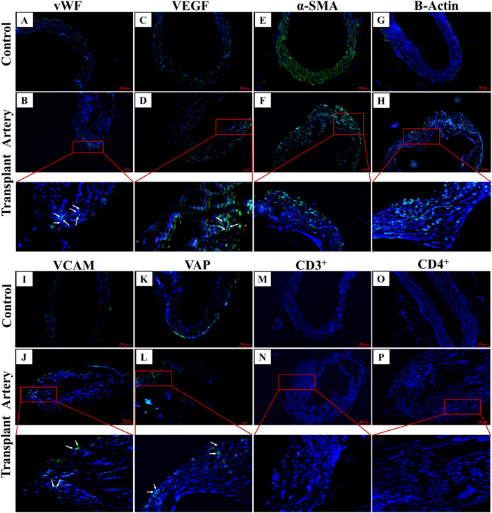
Fig. 7.
(A–P) Immunohistochemistry analysis of transplanted hDA graft done by vWF, VEGF, α-SMA, VCAM, B-Actin, VAP and immunological respose were analysed by CD3 + , CD4 + markers. B Edothelial cells were stained with von Willebrand factor (vWF, green) and F Smooth muscle cells are stained with α-Smooth muscle actin (α-SMA, green). Scale bars: 50 µm

