
Fig. 2.
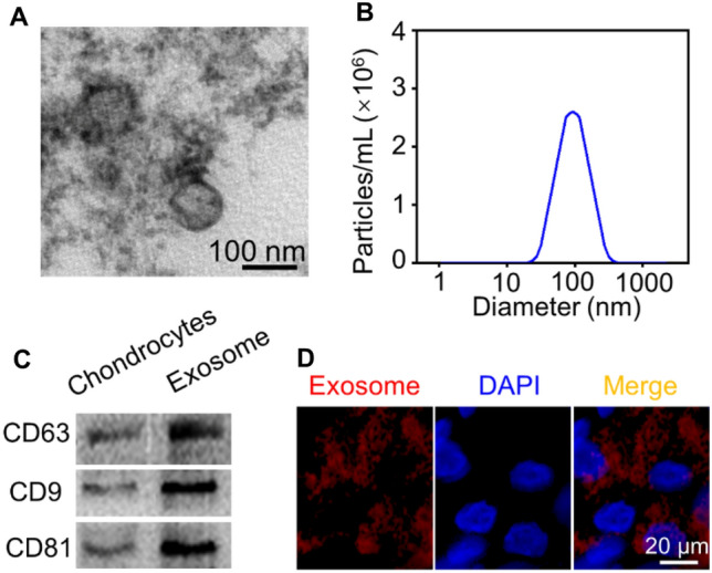
Characterization and uptake of primary chondrocyte derived-exosomes. A TEM images exhibited the morphology of primary chondrocyte derived-exosomes. B NTA detection showed the particle size distribution of exosomes. C Western blotting evaluated the expression of exosome surface markers, CD63, CD81, and CD9. D Representative immunofluorescence images of Dil-labeled exosomes absorbed by chondrocytes