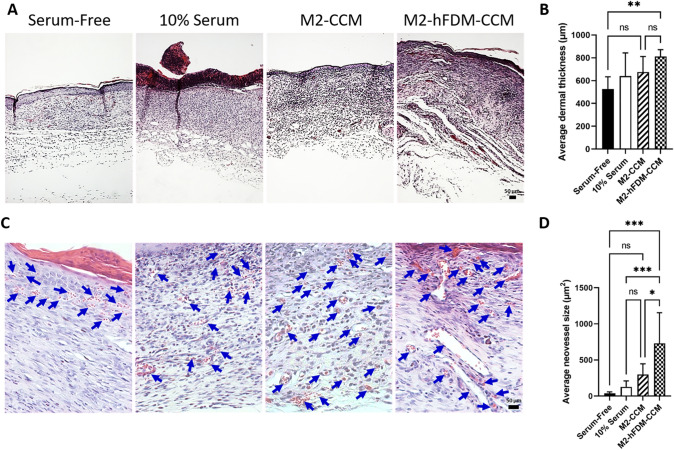
Fig. 6.
CCM-treated wounds reveal better histological outcome and advanced neovascularization. A Representative H&E staining images of the regenerated wound tissues on day 14 post-treatment. B Quantitative analysis of dermis thickness. C High magnification of H&E images shows the neovessels on day 14 post-treatment. Blue arrows mark neovessels. D Quantitative analysis of average vessel size. Scale bar is 50 μm. Statistically significant difference (*p < 0.05, **p < 0.01, ***p < 0.001)

