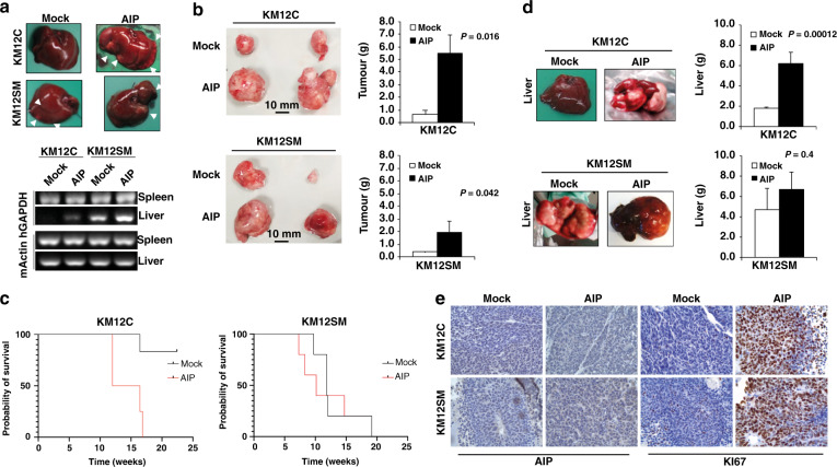
Fig. 5. AIP-ectopic expression induces tumour growth and liver metastasis in KM12 cells.
a Nude mice intrasplenically inoculated with indicated KM12 cells were sacrificed 24 h after inoculation for analysis of in vivo homing. RNA was isolated from the liver and subjected to RT-PCR to amplify human GAPDH (hGAPDH). Representative experiments out of three are shown. Murine β-actin (mβ-actin) was amplified as a control. b KM12 transfectants were inoculated subcutaneously in nude mice (n = 6 per group). Tumour size was measured every day for 4 weeks and mean ± SEM of the endpoint represented by bar graphs. Representative tumour images from KM12 cell stably transfectants are also depicted. Tumours were significantly higher in AIP-transfected cells (P < 0.05) and in AIP-transfected KM12C cells vs KM12SM cells (P = 0.013). c Kaplan–Meier survival assay of nude mice inoculated intrasplenic with the indicated KM12 cell transfectants (n = 5 per group). Survival of mice inoculated with AIP-stably transfected cells significantly decreased (**P < 0.01) when compared with those inoculated with Mock control cells. d Mice were examined for macroscopic metastases in liver. (Left) representative images of macroscopic metastases in the liver significantly induced in mice inoculated intra-spleen with AIP-stably transfected KM12C and KM12SM cells are shown. (Right) Livers were weighed at the endpoint and mean ± SEM represented (n = 5 per group). Livers were significantly heavier in mice inoculated intra-spleen with AIP-stably transfected KM12C and KM12SM cells because of tumour metastasis. e Representative IHC images of FFPE tumours developed in the mice injected with either KM12C or KM12SM Mock or AIP-stably expressing cells. Tumour samples were stained with anti-KI67 or anti-AIP antibodies. A more intense AIP staining was observed in tumours from mice inoculated with AIP-overexpressing cells compared to Mock controls. Likewise, a clearly more intense nuclear KI67 staining was observed in tumours from mice subcutaneously inoculated with AIP-overexpressing cells.

