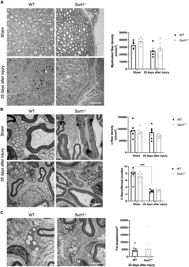
FIGURE 2.
Morphological and morphometrical analysis of crush-injured Sort1–/– sciatic nerve. (A) Myelinated and (B) C-fiber densities were similar between Sort1–/– mice and WT mice in the contralateral (sham) and 25 days after sciatic nerve crush injury. Representative EM images of thin transverse sections of sham contralateral or the distal stumps of crush-injured sciatic nerves 25 days after injury [scale bar (A): 20 μm; (B): 2 μm]. Bottom graph in panel (B) shows the average number of C-fibers/Remak bundle, which were significantly lower (***p < 0.001) in both crush-injured WT and Sort1–/– sciatic nerve compared to the contralateral sham control. (C) Representative images of fat droplets in thin transverse sections of injured WT or Sort1–/– sciatic nerve 25 days after sciatic nerve crush injury. The total number of fat droplets/mm2 were similar between WT and Sort1–/–.

