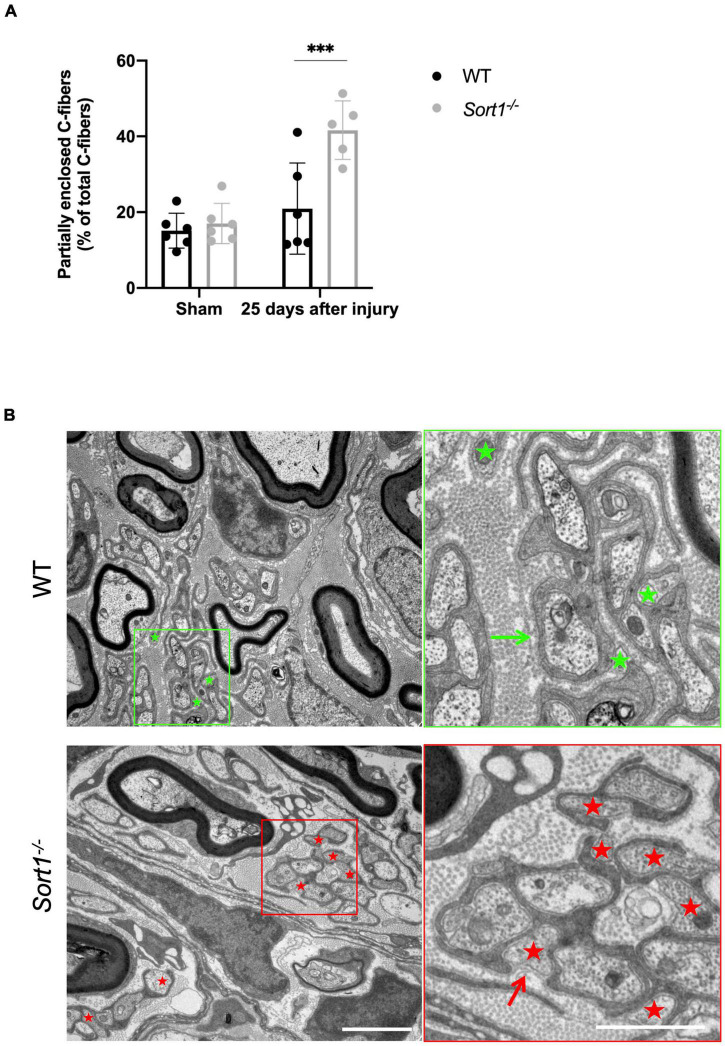
FIGURE 3.
Reduced axon ensheathment by Remak Schwann cells in regenerated Sort1–/– sciatic nerve. Significantly more partially enclosed C-fibers were observed in the injured Sort1–/– sciatic nerve compared to WT. Representative images showing normal Schwann cell ensheathment of C-fibers in injured WT sciatic nerve 25 days after crush injury (indicated by the green arrow) and only partly enclosure of C-fibers by Schwann cell cytoplasm and partly by only basement membrane 25 days after sciatic nerve crush injury in WT (green stars) and the Sort1–/– mouse (indicated by red stars and red arrows). Scale bars: left images 2 μm, right images (zoom of left) 1 μm. (A,B) N = 6 WT mice and 6 Sort1–/– mice. Statistical significance was established with two-way ANOVA and Bonferroni’s multiple comparisons post hoc test (***p < 0.001).

