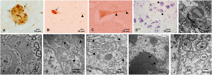
Figure 1.
Immunohistochemistry of hyperphosphorylated tau (p-Tau) in substantia nigrae pars compacta (SNpc) and brainstem, and Electron Microscopy in SNpc from Metropolitan Mexico City subjects. (A) Hyperphosphorylated tau (p-Tau) mature plaque (arrows) in midbrain, 40y old male. (B) Lower medulla p-Tau tangle (short arrow) and a p-Tau positive neurite (arrowhead) in a 13y old girl. (C) Raphe neuron with granular positive p-Tau (arrowheads) staining in a 3y old boy. (D) SNpc, 1μm toluidine blue section showing neurons with abundant cytoplasmic neuromelanin (NM) (arrowheads) in sharp contrast with neurons with scanty cytoplasm with few NM (short arrows). One small vessel (♦) has a vacuolated perivascular neuropil. (E) SNpc neurovascular Unit (NVU). Blood vessels are seen with leaking walls with clusters of lipids in the neuropil. The neuropil is vacuolated (♦). A portion of a macrophage is seen (Mo). (F) 11-month old baby. SNpc neuron with a nucleus (Nu) and significant damage to the surrounding neuropil i.e., large vacuolated spaces with debri and macrophage-like cells (short arrow). (G) 12-year-old, SNpc neurons with dilated endoplasmic reticulum ER (arrowheads), nucleus marked (♦). (H) 26 year old, SNpc dilated ER (arrowheads). (I) Nanoparticles inside a SNpc neuronal nucleus (arrowheads) (J) SNpc abnormal mitochondria (M) and dilated ER.

