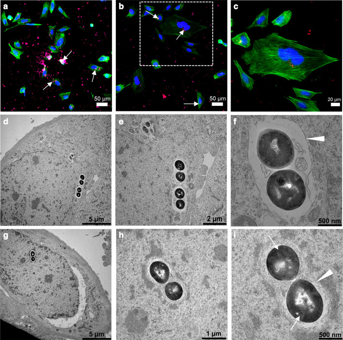
Fig. 1.
Intracellular invasion of Staphylococcus aureus in osteoblasts. a) to c) Immunofluorescence images showing clusters of S. aureus EDCC 5055 that are localized inside the Saos-2 cells at three hours post-infection. The S. aureus (white arrows), nuclei, and actin filament are stained in red, blue, and green, respectively. d) to i) Ultrastructural imaging of intracellular S. aureus EDCC 5055 revealed internalized bacteria clusters localized close to the cell nuclei membrane. Most bacteria clusters possess intact cell wall/plasma membrane and are surrounded by phagosomal membrane (white arrowheads). Some proliferating bacteria revealed division septa (white arrows).

