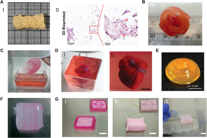
FIGURE 5.
The applications of ECM-based bioinks. (A) (I) A clavicle bone scaffold bioprinted with BPs GelMA-based bioink. (II) The scaffold stained for H&Eafter 28 days, the number of cells increased. Adapted with permission from (Ratheesh et al., 2020). (B) The aortic valve conduit bioprinted with bioink containing alginate/gelatin hydrogel and aortic root sinus smooth muscle cells and aortic valve leaflet interstitial cells. Adapted with permission from (Duan et al., 2013). (C) Adult size ears (8 cm) printed with bioink containing bovine gelatin/alginate/fibrinogen and human fibroblasts. Adapted with permission from (Pourchet et al., 2017). (D) A heart printed with bioink containing dECM and iPSCs-derived cardoimyocytes and ECs. Adapted with permission from (Noor et al., 2019). (E) The curved cornea based on the eyeball printed with dECM-based bioink. Adapted with permission from (Kim H. et al., 2021). (F) The multilevel vascular structures, and (G) multibranch vascular channels printed with bioink containing dECM/Pluronic F127 and endothelial cells. Adapted with permission from © 2018 by the (Xu et al., 2018). Licensee MDPI, Basel, Switzerland.

