Figure 1.
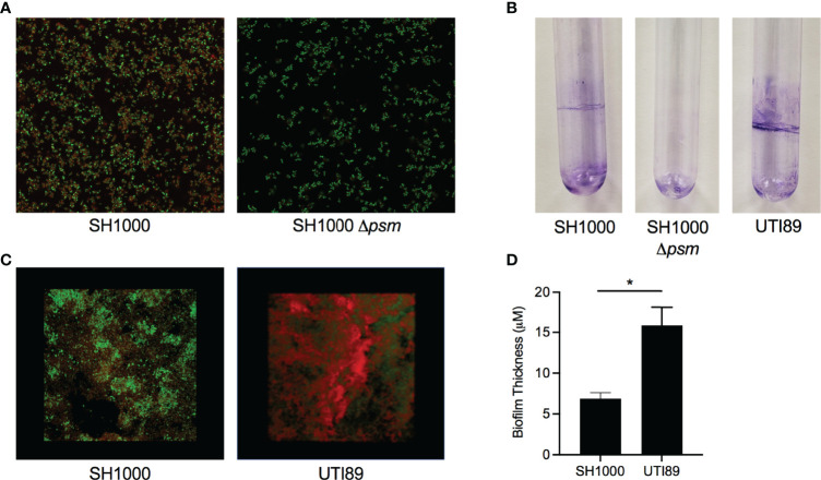
Staphylococcus aureus biofilms contain amyloid that can be detected by an amyloid specific stain. (A) Confocal Laser Scanning Microscopy (CLSM) images of in vitro biofilms of S. aureus SH1000 (lab strain) and SH1000 phenol soluble modulin mutant (Δpsm) were stained with syto9 (green) and FSB (red) to determine the expression of PSM amyloids. (B) Crystal Violet staining of pellicle biofilms grown in glass tubes of SH1000, SH1000 Δpsm, and UTI89 (E. coli clinical isolate). (C) CLSM surface images of in vitro biofilms of S. aureus SH1000 and E coli UTI89 were stained with syto9 (green) and FSB (red). (D) Overall biofilm thickness of SH1000 and UTI89 biofilms as determined by Leica TCS imaging software. Mean and SEM graphed; significance was calculated using Unpaired t test (*, P < 0.05).

