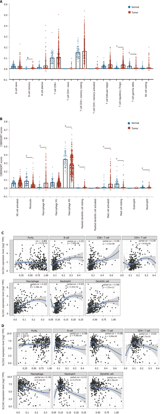
Figure 7.
The landscape of infiltrating immune cells in hepatocellular carcinoma was different from that in normal liver tissues and expressions of solute carrier family 2 member 1 and solute carrier family 2 member 2 correlated with immune infiltration level in hepatocellular carcinoma. A and B: The pattern of immune cells by the signature gene expression profile in hepatocellular carcinoma compared with normal samples with the Cell Type Identification by Estimating Relative RNA Transcript subsets method. In comparison to normal tissues, the proportions of B cell memory, regulatory T, T cell gamma delta, monocyte, macrophage M0, macrophage M2, myeloid dendritic cell resting, mast cell activated, mast cell resting, and neutrophil had changed; C: Correlation analysis between solute carrier family 2 member 1 (SLC2A1) transcription level and immune cell infiltration level. The immune cells included B cells (partial.cor (r) = 0.291), CD8+ T cells (r = 0.296), CD4+ T cells (r = 0.238), macrophages (r = 0.425), neutrophils (r = 0.424), and dendritic cells (r = 0.368); D: Correlation analysis between solute carrier family 2 member 1 (SLC2A2) transcription level and immune cell infiltration level. The immune cells included B cells (r = -0.188), CD8+ T cells (r = -0.188), macrophages (r = -0.148), neutrophils (r = -0.115) and dendritic cells (r = -0.17). aP < 0.05; cP < 0.001.

