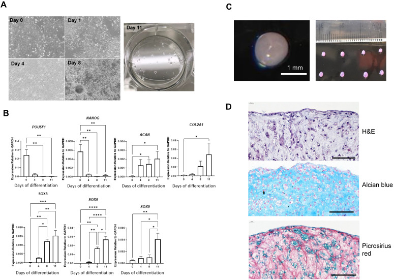
Figure 2.
Differentiation of MAN13 stem cells into pre-chondrocytes through the RAPID protocol. (A). Cell morphology showing MAN13 hESCs at day 1, 4, 8 and 11 with the appearance of cell aggregates. (B). qPCR analysis of pluripotency-associated and chondrogenic marker genes during the 2D stage (11 days) of the RAPID differentiation protocol. N = 3-5. *, P < 0.05; **, P < 0.01; ***, P < 0.001; ****, P < 0.0001. (C). Photographs of 3D chondrogenic pellets. (D). Histology evaluation of 3D pellets (Haematoxylin and Eosin, Alcian blue and Picrosirius red). N = 3, scale bars = 100 µm.

