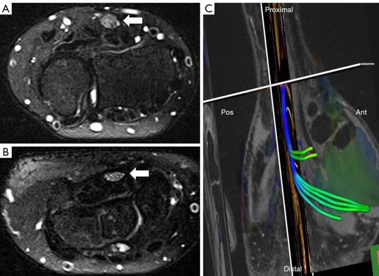
Figure 1.
MRI of median nerve at the carpal tunnel. (A) Median nerve (arrow) at the distal radioulnar joint. (B) Median nerve (arrow) at the outlet of the carpal tunnel in a patient with CTS. (A) and (B) axial T2-weighted spectral attenuated inversion recovery. (C) Reconstructed tractography of the median nerve through the carpal tunnel. MRI, magnetic resonance imaging; CTS, carpal tunnel syndrome.

