Figure 1.

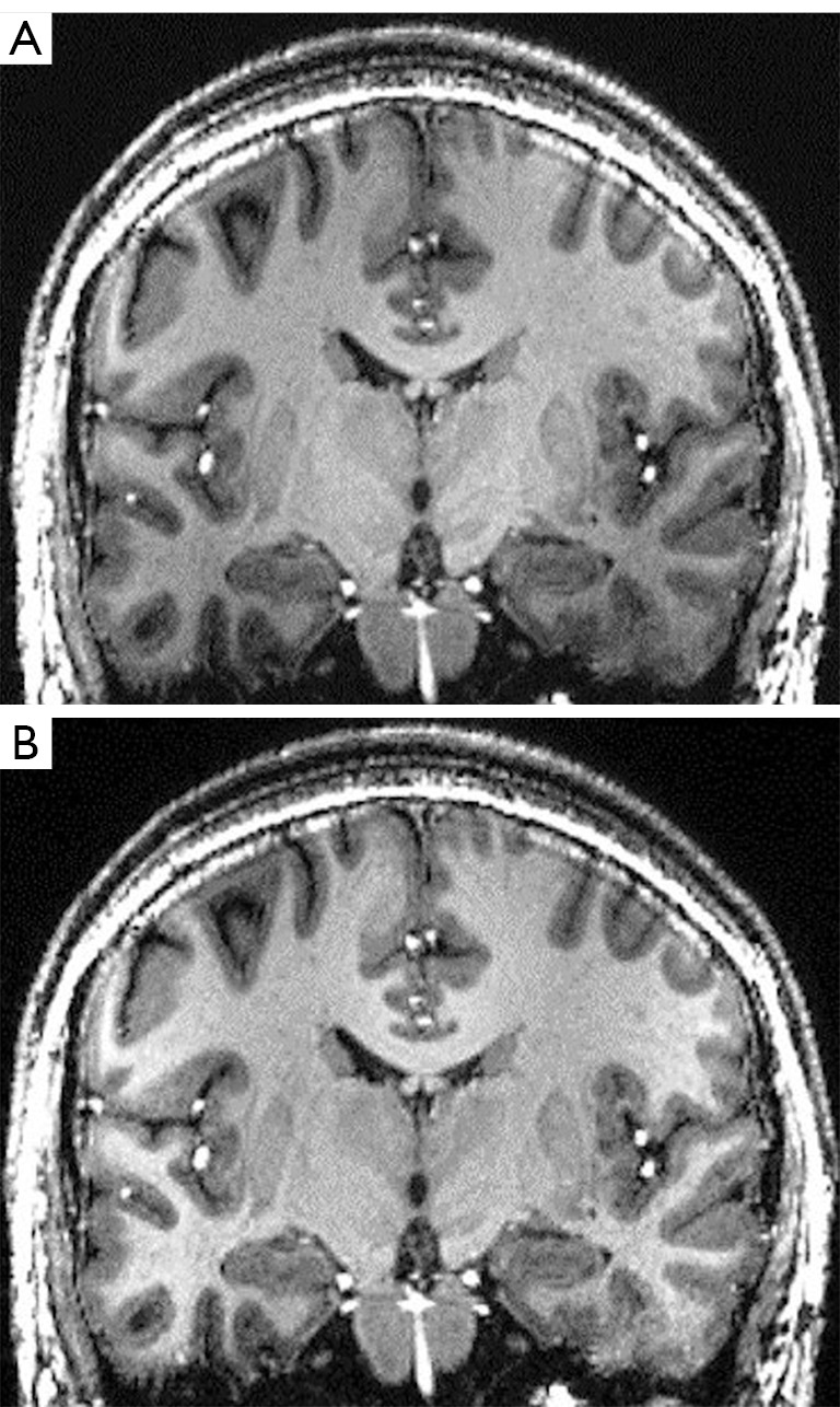
T1-weighted coronal MPRAGE images before (A) and after (B) signal inhomogeneity correction. Gradual signal decrease toward the skull base is corrected. MPRAGE, magnetization-prepared rapid gradient echo.

T1-weighted coronal MPRAGE images before (A) and after (B) signal inhomogeneity correction. Gradual signal decrease toward the skull base is corrected. MPRAGE, magnetization-prepared rapid gradient echo.