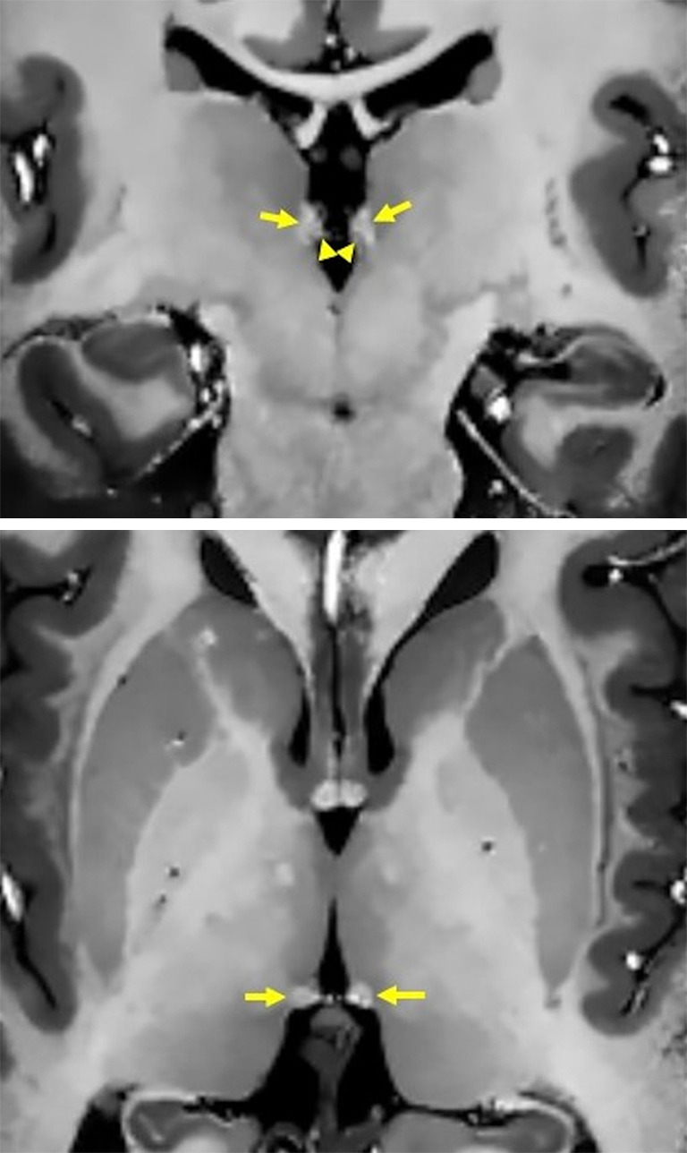
Figure 10.

High-resolution T1WI (0.5 mm isotropic resolution) of the habenula (arrows) acquired using MP2RAGE after denoising (top: coronal, bottom: axial). On the coronal image, the lateral nucleus shows a slightly higher signal reflecting a shorter T1 value than that of the medial nucleus (arrowheads). T1WI, T1-weighted imaging; MP2RAGE, magnetization-prepared 2 rapid gradient echoes.
