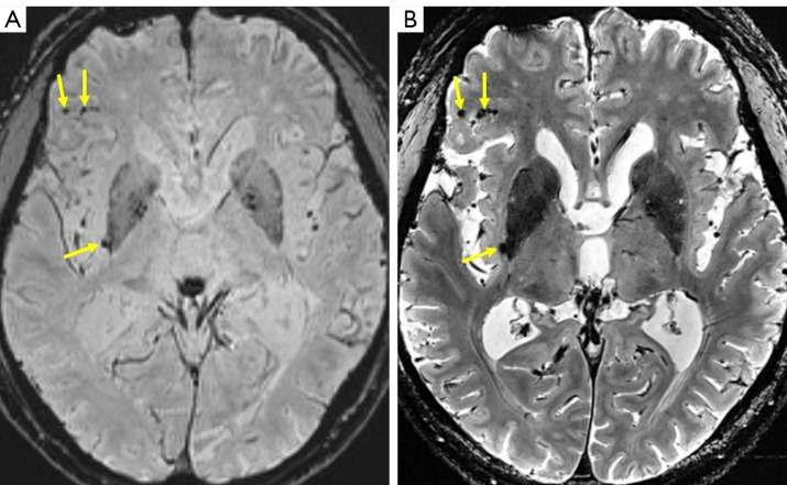
Figure 2.
A patient with a traumatic brain injury. (A) SWI at 3T shows small hemorrhagic lesions as low-intensity spots (arrows), but their relation to the background structure is relatively obscured, and their locations in the cortex or sulci are ambiguous. (B) T2*WI at 7T enables easy detection of small hemorrhagic spots (arrows), including their anatomical location. SWI, susceptibility-weighted imaging; T2*WI, T2*-weighted imaging; 3T, 3 Tesla; 7T, 7 Tesla.

