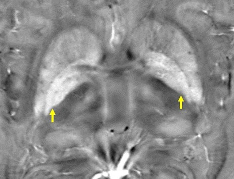
Figure 9.

An axial QSM image of a healthy subject at the basal ganglia. The globus pallidus interna is separated from the externa by the medial medullary lamina (arrows), which is visualized as a thin layer of low signal intensity. Differences in susceptibility can also be observed among the thalamic subnuclei. QSM, quantitative susceptibility mapping.
