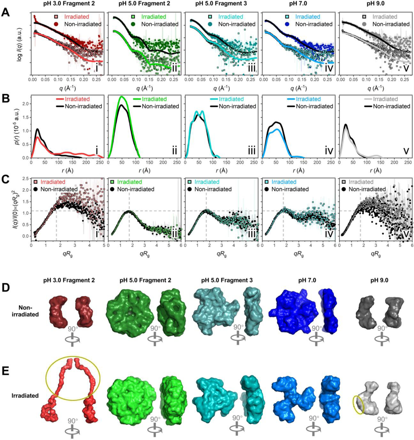
Figure 4. SAXS analysis reveals irradiation-induced conformational changes of C-PC at pH 3.0–9.0.

(A) SAXS scattering profiles (dots) fitted with theoretical scattering curves (lines) back-calculated from corresponding C-PC crystal models (PDB 1GH0).11 Non-irradiated fragments are colored lines on square dots; irradiated fragments are black lines on circular dots. The following C-PC crystal models in different assembly states were used for fitting: (αβ) monomer in i, v; (αβ)3 trimer in iii, iv; and (αβ)6 hexamer in ii. (B) Dimensionless Kratky plots. (C) Pair distance distribution functions. (D–E) Surface representations of GASBOR ab initio envelopes for non-irradiated (D) and irradiated (E) C-PC fragments. For Fragment 2 at pH 3.0 (red) and the fragment at pH 9.0 (grey), the dotted ovals in (E) highlight the pronounced protrusions that arised in the structures after irradiation, which were absent from the corresponding non-irradiated structures in (D). The corresponding DAMMIN envelopes in Figure S6, E and F, reveal similar protrusions in the structures.
