
Figure 1: Spatiotemporal control over organoid crypt formation through photopatterning.
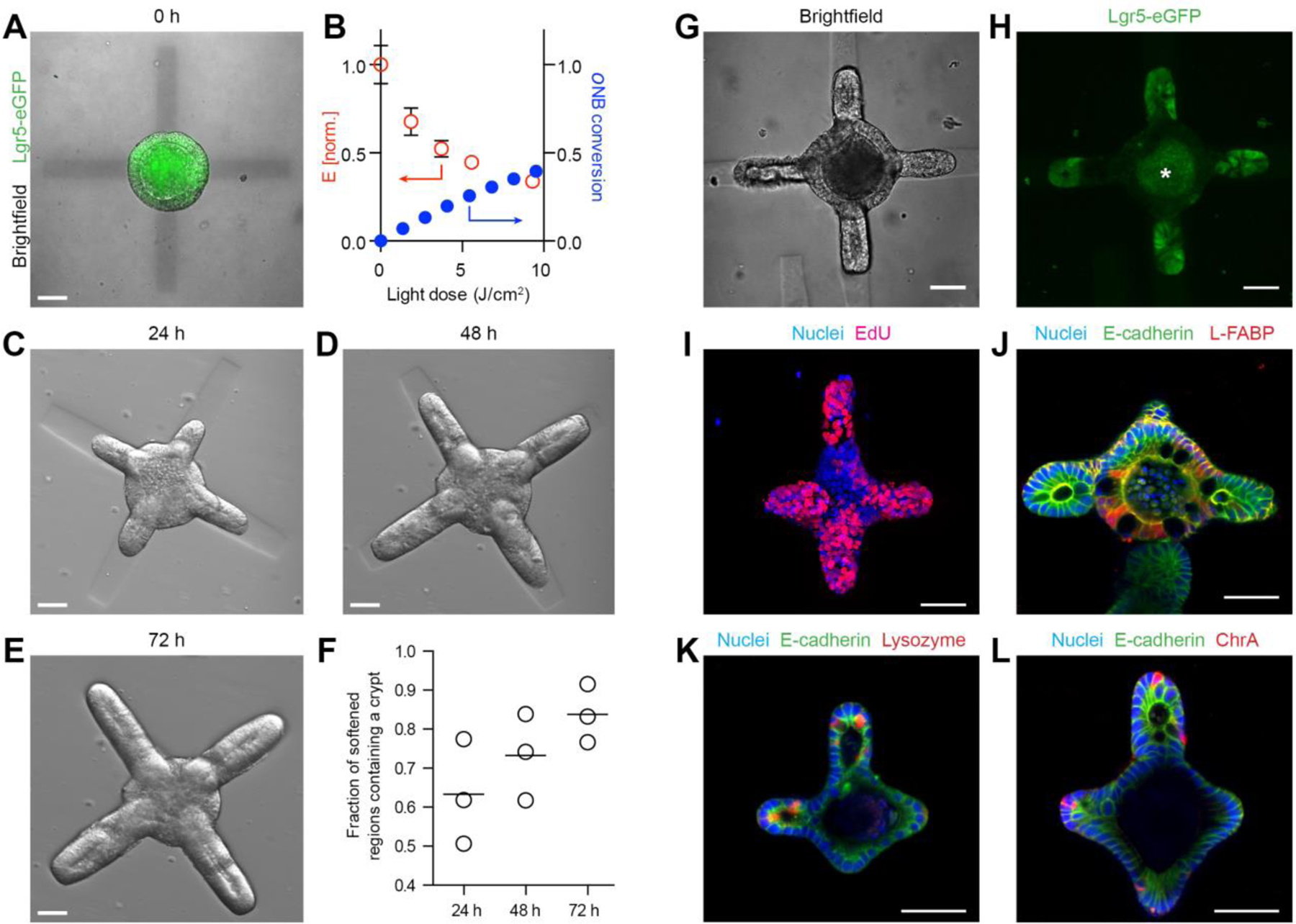
(A) Composite image showing Lgr5-GFP expression in a symmetric colony and photopattern visible with transmitted 405 nm light immediately after spatially restricted light exposure. (B) Mechanical characterization of gel regions with atomic force microscopy corresponds to conversion of photocleavable moieties within the gel. (C-E) Spatially defined crypt formation within photopatterned gels 24 h (C), 48 h (D) and 72 h (E) after light-induced softening. (F) Quantification of fraction of photo-softened gel regions containing a crypt. Individual data points and mean are shown. Lgr5-eGFP expression (G, H) and proliferation (I) are localized within the buds extending into the softened regions. Enterocytes are found in the central regions (J) of the organoids Paneth cells are restricted to the buds (K) of the organoids. Enteroendocrine cells (L) are also present. Scale bars, 30 µm.