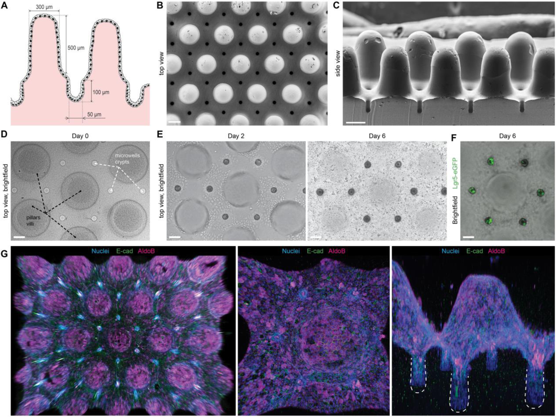
Figure 4: Bioengineered organoids with an in vivo-like tissue architecture.

(A) Scheme of the designed topography resembling the native tissue with characteristic intestinal crypt-villus architecture. (B, C) SEM images of the poly(dimethylsiloxane) (PDMS) template used for fabricating bioengineered hydrogel substrates featuring a crypt-villus architecture, top and side view. Scale bars, 200 μm. (D) Top view of the hydrogel substrate shaped according to the topology of the native intestinal mucosa. (E) Brightfield time-course images of the intestinal epithelium development. (D,E) Extended depth of field for a z-stack ~600 μm. (F) Localization of the Lgr5+ stem cells in the engineered crypts. (D-F) Scale bars, 100 μm. (G) 3D reconstruction of the immunofluorescence images showing confluent monolayer of E-Cadherin expressing epithelial cells covering hydrogel substrates, harboring villi composed of enterocytes (AldoB) and other differentiated cell types.
