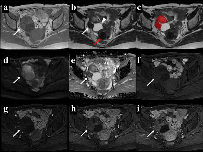
Fig. 2.
A 66-year-old woman with right ovarian fibrothecoma. Axial T1WI (a) showed an isointense solid mass and axial T2WI (b) showed a heterogeneous solid mass with a cystic change in the dorsal part (arrowhead). Uterine fibroids were present (red arrow). The region of interest (ROI) in the texture analysis (c) was delineated to contain the outline borders of the solid region of the tumor, and the cystic region was avoided as much as possible. Axial DWI (d) showed a hyperintense solid area, and the apparent diffusion coefficient (ADC) map (e) showed that the average ADC value of this area was 1.31 × 10⁻³ mm2/s. In multiphasic contrast-enhanced fat-suppressed T1-weighted MRI (f-i), the mass showed minimal enhancement in the arterial and venous periods (g and h, respectively) as well as heterogeneous mild enhancement in the delayed period (i)

