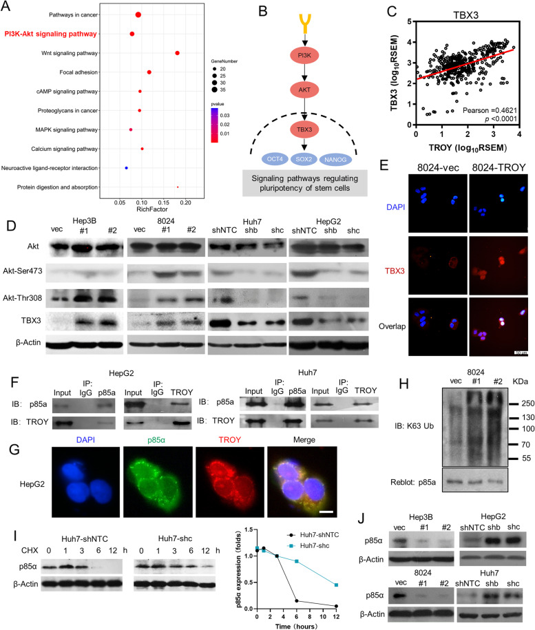
Fig. 5.
TROY activates PI3K/AKT/TBX3 signaling pathway via polyubiquitin p85a. (A) KEGG pathway enrichment study of 823 up-regulated genes in TROYhi HCC patients. (B) PI3K/AKT/TBX3 signaling pathway from Signaling pathways regulating pluripotency of stem cells21. (C) Co-expression analysis of TROY and TBX3 from TCGA datasets. The R-value was detected by Pearson correlation, the P-value was tested by independent-samples t-test. (D) Western blotting of Akt, Phospho-Akt (Ser473 and Thr308), and TBX3 in TROY overexpression and knockdown HCC cells. β-Actin was used as a loading control. (E) Representative immunofluorescence images of TBX3 in 8024 transduced with vector or TROY. Scale bar = 50 μm. (F) Cell lysates prepared from HepG2 and Huh7 were subjected to immunoprecipitation (IP) with TROY antibody or control immunoglobulin G (IgG) and then immunoblotted with p85α antibody. (G) IF double staining of TROY and p85α in HepG2 cells. Nuclei were stained with DAPI. Scale bar, 20 μm. (H) IP with anti-p85α antibody and blotted with anti-k64 antibody in 8024 cells transfected with vec or TROY isoforms. (I) Western blotting (Left) and quantification (right) of ShNTC- or shTROY-Huh7 cells incubated with CHX (40 μmol/L) for the indicated time points. (J) Western blotting of p85α in TROY overexpression and knockdown HCC cells. β-Actin was used as a loading control. Statistical significances: ***, P < 0.001

