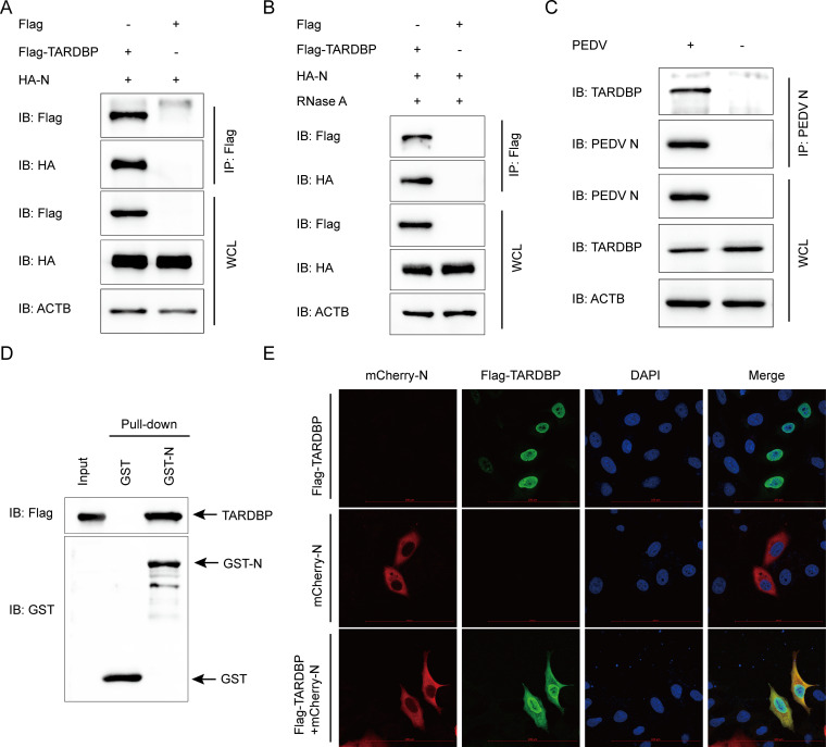
FIG 3.
TARDBP can interact with PEDV N protein. (A) An entire day of transfection of HEK 293T cells was accomplished using the HA-N- and Flag-TARDBP-encoding plasmids, and then the anti-Flag binding beads were utilized for the co-IP procedure. Western blotting was used for analyzing the precipitated proteins. ACTB was applied as a control. WCL, whole-cell lysate. (B) The interaction of TARDBP with PEDV N protein after RNase treatment. (C) Following pseudoinfection or infection using PEDV at an MOI of 0.01, the Vero cells were gathered for the endogenous TARDBP immunoprecipitation based on the antibody of N protein. (D) The pCold TF and pCold GST plasmids were utilized for independent cloning of TARDBP and PEDV N, which were subsequently denoted in BL21(DE3) bacterial strain for the affinity isolation of GST. The eluted proteins were explored with the use of Western blotting. (E) Following an entire day of transfection of HeLa cells using N-mCherry- and Flag-TARDBP-encoding plasmids, the specific antibodies were utilized to accomplish cellular labeling. DAPI labeling was used for the cellular nuclei, while a confocal immunofluorescence microscope was utilized for the monitoring of fluorescent signals (scale bars, 100 μm).

