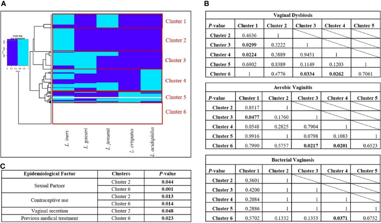
Figure 3.
Clustering of the vaginal samples according to the prevalence of Lactobacillus sp. (A) Clusters obtained by Ward’s Minimum Variance Clustering Method. (B) Chi-square analysis between clusters in presence of vaginal dysbiosis, aerobic vaginitis, and bacterial vaginosis. (C) Epidemiological factors related to each cluster. Six clusters were chosen according to the presence of different Lactobacillus species in vaginal samples using Ward’s Minimum Variance Clustering Method. The following clusters are in panel (A), Cluster 1 was characterized by the presence of Lactobacillus iners and Lactobacillus jensenii; Cluster 2 only showed L. iners; Cluster 3 was constituted by L. iners, L. jensenii, and Lactobacillus gasseri; Cluster 4 was formed by L. iners, L. jensenii, L. gasseri, and Lactobacillus acidophilus; Cluster 5 is a mixture of all Lactobacillus species; and Cluster 6 shows the absence of all of them. The dark blue color indicates the absence of a Lactobacillus species; meanwhile, the light blue color indicates the presence of the Lactobacillus species. In panel (B), chi-square tests were performed to assess the statistical differences between clusters. The p-values where statistically significant differences were found are shown in bold. Finally, in panel (C), multiple chi-square tests were performed to evaluate the epidemiological factors related to the presence of each cluster; the significant values are featured in the corresponding table in bold.

