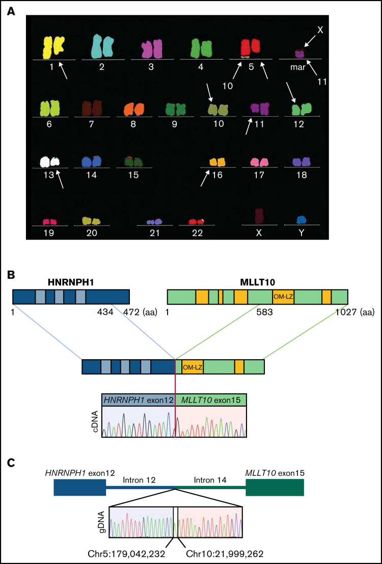
Figure 1.
Molecular characterization of the HNRNPH1-MLLT10 fusion. (A) Multicolor fluorescence in situ hybridization indicating a complex karyotype including del(5q). (B) HNRNPH1-MLLT10 splicing isoform identified in the present case is shown. GenBank accession numbers referred to are NM_001364227.2 for HNRNPH1 and NM_004641.4 for MLLT10. Hypothetical fusion protein is shown in which HNRNPH1 maintained all 3 RNA-recognition motifs at the N-terminus and MLLT10 maintained the critical OM-LZ domain at the C-terminus. Sanger sequencing of complementary DNA revealed that exon 10 of HNRNPH1 (rather than exon 12, which was reported previously) fused in frame with exon 15 of MLLT10 (as reported previously). (C) Sanger sequencing indicates genomic breakpoints at position Chr5:179 042,232 within HNRNPH1 gene and Chr10: 21 999,262 in MLLT10 gene with a single nucleotide insertion.

