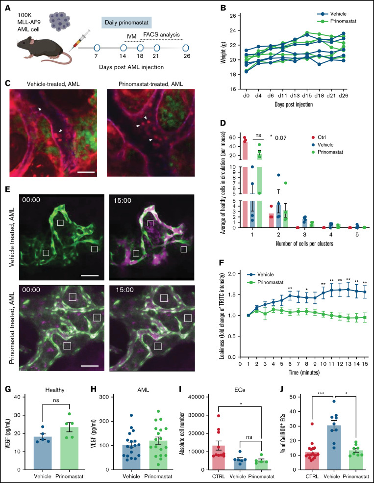
Figure 2.
Prinomastat reduces circulating healthy cell clusters and rescues vascular leakiness. (A) Schematic of the prinomastat treatment regime adopted and timing of IVM and flow cytometry analyses. (B) Total body weights of vehicle and prinomastat-treated AML-burdened mice throughout the experiment. (C) Selected, representative timeframes of time-lapse IVM showing YFP+ AML blasts (green) and circulating mTomato+ healthy hematopoietic cells (red) in vehicle- and prinomastat-treated leukemic mice. Blood vessels are stained with Alexa647 anti-VE–cadherin antibody, resulting in purple staining of tomato+ endothelial cells. Arrowheads point at clusters of healthy cells in vehicle-treated and single cells in prinomastat-treated leukemic mice, respectively. Scale bar represents 25 µm. (D) Quantification of circulating healthy single cells and clusters in vehicle- and prinomastat-treated leukemic mice. Shown are average numbers of cells/clusters detected per FoW in 3–hour-long (3 min time frame) time-lapse datasets. *P < .05. Data pooled from 11 mice in total from 3 independent experiments. (E) Vascular leakiness was assessed by time-lapse IVM of randomly selected regions within the calvarium of leukemic Flk1-GFP mice following administration of TRITC-dextran IV. Selected representative frames from vehicle- and prinomastat-treated leukemic mice (n = 6 mice/condition) are shown. AML cells are not shown. Scale bar represents 80 µm. (F) Quantification of the fold change in TRITC-dextran intensity in the BM parenchyma of vehicle- and prinomastat-treated mice. n = 6 vehicle- and n = 6 prinomastat-treated, leukemic mice. Three areas measured/mice (eg, white boxes in [E]). (G) VEGF levels (pg/mL) measured by ELISA in the BM supernatant of healthy (n = 5 mice per condition) and (H) leukemic mice treated with vehicle or prinomastat. (I) Quantification of the absolute number of endothelial cells in healthy control and vehicle and prinomastat treated AML-burdened mice. Data pooled from 21 mice from 2 independent experiments. (J) Quantification of CellROX+ endothelial cells in healthy control and vehicle and prinomastat-treated mice. n = 8 per condition. All data are mean ± SEM. *P < .05; **P < .01; ***P < .001; ns, not significant. P values determined by multiple Student t tests with posthoc Holm-Sidak corrections.

