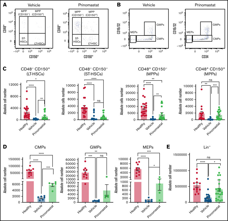
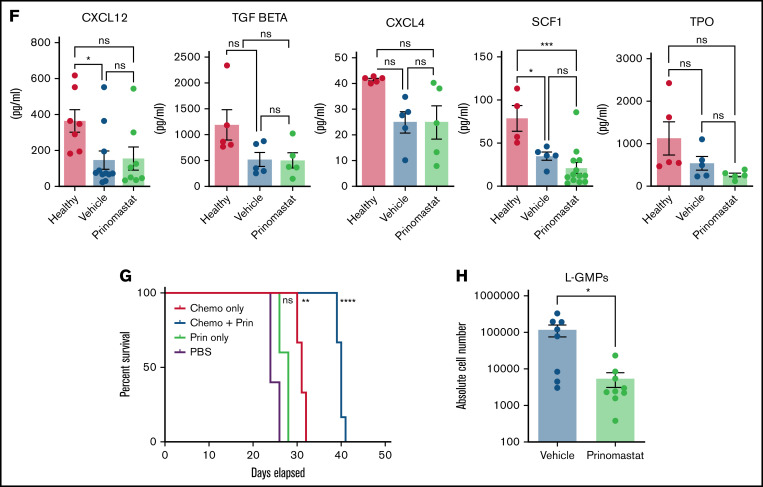
Figure 5.
Prinomastat protects healthy HSPCs and, combined with chemotherapy, increases survival. (A-B) Flow cytometry plots showing LKS and Lin- cKit+ Sca-1- cell populations in leukemic mice that received PBS or prinomastat, and with >70% BM infiltration. (C) Absolute number of LT-HSCs, ST-HSCs, and MPP populations per leg in healthy and leukemic, PBS- or prinomastat-treated mice. (D) Absolute number of CMPs, GMPs, MEPs per leg in healthy and leukemic, PBS- or prinomastat-treated mice. (E) Absolute number of lineage- cells per leg in healthy and leukemic, PBS- or prinomastat-treated mice. (C-E) n = 5-22 mice per condition pooled from up to 5 independent experiments. (F) CXCL12, TGF-β, CXCL4, SCF1, TPO levels (pg/mL) measured in BM supernatant from healthy control, vehicle- and prinomastat-treated leukemic mice by ELISA. Each dot represents 1 mouse. n = ≥3 per condition for each of the tested factors. (G) Kaplan-Meyer curve showing survival of mice receiving chemotherapy alone, prinomastat alone, prinomastat in combination with chemotherapy, or vehicle control (PBS). n = 6 for chemotherapy only and chemotherapy + prinomastat groups. n = 5 for prinomastat and PBS only groups. (H) Absolute number of L-GMPs within the BM of vehicle- and prinomastat-treated leukemic mice (day 23). n = 8 vehicle-treated and n = 9 prinomastat-treated AML burdened mice. Data are shown as mean ± SEM. *P < .05; **P < .01; ***P < .001; ****P < .0001; ns, not significant. P values determined using multiple Student t tests with posthoc Holm-Sidak corrections or 1-way ANOVA and the log-rank test in (G).


