. 2022 Apr 26;13(20):6019–6027. doi: 10.1039/d2sc00918h

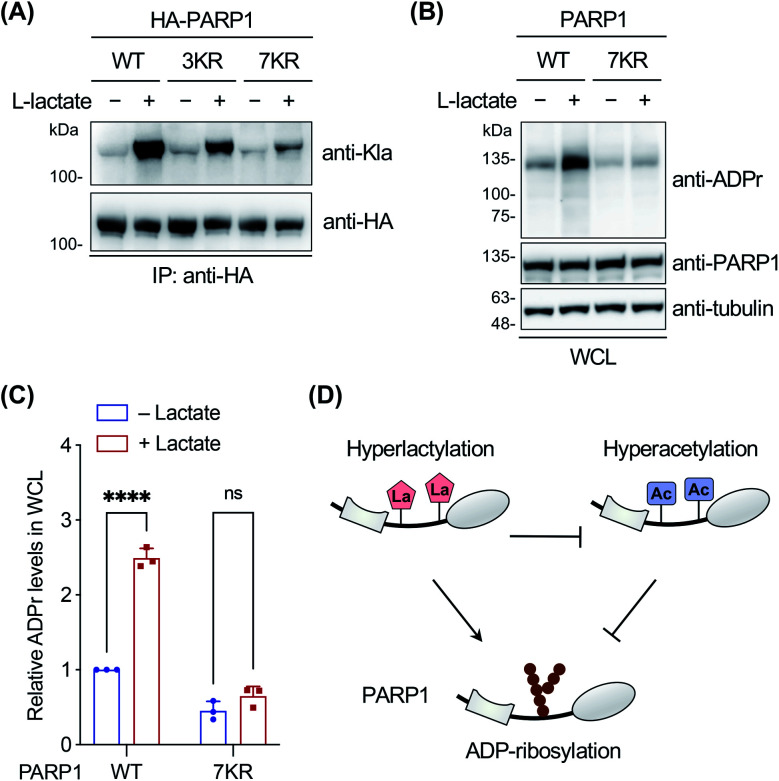
Fig. 5. Investigation on PARP1 lactylation. (A) Analysis of lactylation of PARP1, 3KR (K498/505/506R), and 7KR (K498/505/506/508/518/521/524R) mutants. HEK293T cells expressing HA-tagged PARP1 or mutants were treated with or without sodium l-lactate (25 mM) and lysed for immunoprecipitation. The immunoprecipitates were analyzed by western blot using a pan anti-lactyllysine antibody (anti-Kla). Anti-HA blotting shows the protein loading for each lane. (B) Analysis of ADP-ribosylation activity of wild-type (WT) PARP1 and the 7KR (K498/505/506/508/518/521/524R) mutant. PARP1 knockout HEK293T cells were transfected with PARP1 or the 7KR mutant, treated with or without sodium l-lactate (10 mM), and stimulated with 2 mM H2O2 for 10 min. Whole cell lysates (WCL) were prepared for western blot analysis using an anti-ADP-ribosylation antibody (anti-ADPr). (C) Quantification of overall ADP-ribosylation levels relative to anti-PARP1 signals shown in (B). Data are represented as mean ± s.d., n = 3. **** indicates a p-value < 0.0001, and ns indicates a p-value > 0.05, calculated by two-way ANOVA test. (D) Model for differential regulation of PARP1 ADP-ribosylation by hyperlactylation and hyperacetylation in the auto-modification domain.

Fig. 5