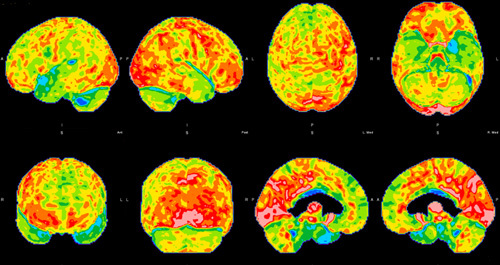
FIGURE 1.

Brain 18fluorodeoxyglucose-positron emission tomography hypometabolism of the first patient. Hypometabolism can be seen in the left temporal lobe, in particular superior temporal gyrus, and mild hypometabolism in the adjacent left frontal and parietal regions with mildly asymmetric decreased tracer activity in the left basal ganglia and thalamus.
