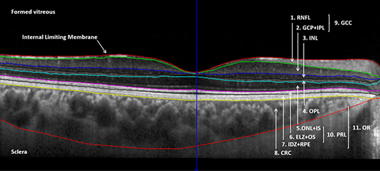
Fig 2. Segmented macular OCT image showing all segmented boundaries by the OCTRIMA 3D algorithm.
The segmented retinal layers shown on the image are the following: retinal nerve fiber layer (RNFL), ganglion cell and inner plexiform layer complex (GCL+IPL), inner nuclear layer (INL), outer plexiform layer (OPL), complex layer containing the Henle fiber layer, outer nuclear layer, external limiting membrane and the myoid zone of the photoreceptors (ONL+IS), complex layer containing the ellipsoid zone and the outer segment of the photoreceptors (ELZ+OS), complex layer containing the interdigitation zone, retinal pigment epithelium and Bruch’s complex (IDZ+RPE) and choroid containing the choriocapillaris, Sattler’s layer and Haller’s layer as far as the choroidal-scleral juncture (CRC). Composite layers, such as the ganglion cell complex (GCC), photoreceptor layer (PRL) and outer retina (OR) are also shown in the figure.

