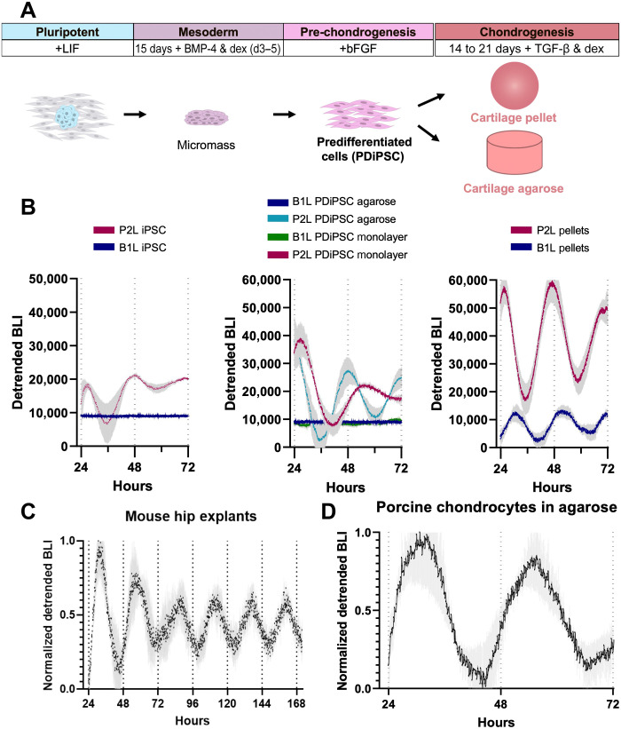
Fig. 2. Development of the circadian clock in miPSC chondrogenesis.
(A) Differentiation protocol of miPSCs to chondrocytes. miPSCs were subjected to high-density micromass culture to create PDiPSCs. PDiPSCs were then either cast in agarose or pelleted and cultured in chondrogenic media for 14 to 21 days to create tissue-engineered cartilage. (B) P2L bioluminescence intensity (BLI) of miPSCs (n = 10), PDiPSCs (n = 2), PDiPSCs in agarose (n = 4), and pellets (n = 10) (left) and B1L bioluminescence intensity (BLI) of miPSCs (n = 5), PDiPSCs (n = 4), PDiPSCs in agarose (n = 5) (middle), and pellets (n = 5) (right). (C) BLI of femoral head cartilage explants from PER2::LUC mice (n = 3, one hip per mouse). (D) P2L BLI in porcine chondrocytes cast in agarose (n = 5). Shaded region on graphs represents SEM.

