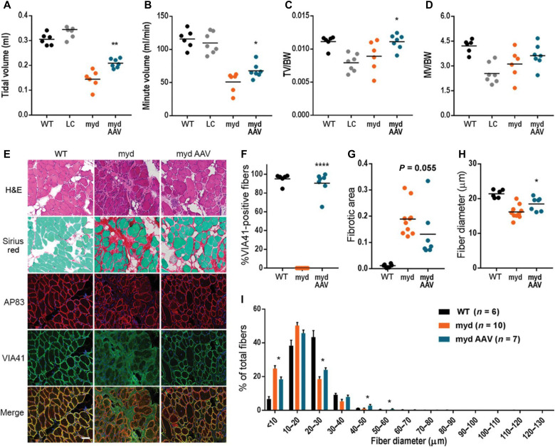
Fig. 3. Large1 gene transfer improves respiratory function and diaphragm pathology.
Whole-body plethysmography for (A) TV, (B) MV, (C) TV normalized to body weight (TV/BW), and (D) MV normalized to weight (MV/BW). (E) Representative cryosections. Sections stained with H&E or Sirius red and Fast Green, or used for immunofluorescence: β-DG (AP83) and matriglycan-positive α-DG (VIA41). Scale bar, 50 μm. (F to I) Quantitative analysis of sections in (E). (F) VIA41-positive fibers. (G) Connective tissue deposition. (H) Average Feret’s diameter of fibers. (I) Percentage of fibers of indicated diameter. Symbols, individual mice; bars, means ± SEM. WT, C57BL/6J; myd, untreated myd; myd AAV, AAVLarge1-injected myd mice; LC, littermate control (Large+/+ or Large+/-).

