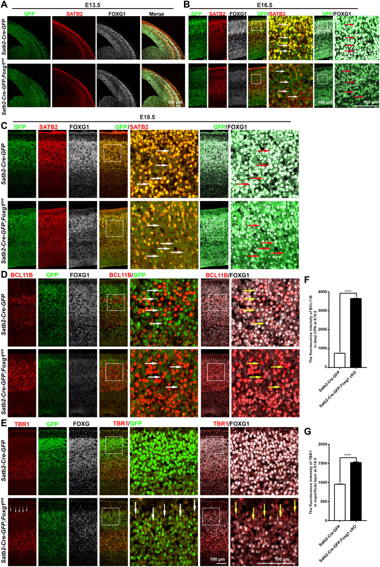
Fig. 8. Disruption of Foxg1 in CPNs.
(A) Triple immunostaining for GFP, SATB2, and FOXG1 at the E13.5 cortex, showing that very few GFP+SATB2+ CPNs existed in the CP in both control and Satb2-Cre-GFP;Foxg1 cKO mice. The level of FOXG1 was comparable between control and Satb2-Cre-GFP;Foxg1 cKO mice. SATB2 level was also comparable. (B and C) Triple immunostaining for GFP, SATB2, and FOXG1 at the E16.5 and E18.5 cortices, showing that GFP was strongly coexpressed in both of SATB2+ deep CPNs and superficial CPNs (arrows) in control mice. In Satb2-Cre-GFP;Foxg1 cKO mice, the levels of both FOXG1 and GFP were significantly decreased, and SATB2 was also severely reduced in GFPweakFOXG1weak neurons (arrows). (D) Triple immunostaining for GFP, BCL11B, and FOXG1 at the E18.5 cortex. BCL11B was not expressed in many GFPstrongFOXG1strong deep CPNs in the control mice (arrows). In the Satb2-Cre-GFP;Foxg1 cKO mice, BCl11B significantly accumulated in GFPweakFOXG1weak neurons in the deep layer (arrows). (E) Triple immunostaining for GFP, TBR1, and FOXG1 at the E18.5 cortex, showing a slight increase in TBR1 in GFPweakFOXG1weak neurons in the superficial layer in the Satb2-Cre-GFP;Foxg1 cKO mice (arrows). (F) Quantification of fluorescence intensity, showing that BCl11B was significantly increased in GFPweakFOXG1weak neurons of the deep layer in the Satb2-Cre-GFP;Foxg1 cKO mice. (G) Quantification of fluorescence intensity, showing that TBR1 level was increased in GFPweakFOXG1weak neurons, which populated in the superficial layer. Data were presented as means ± SEM; unpaired Student’s t test. ****P < 0.0001.

