
Figure 1.
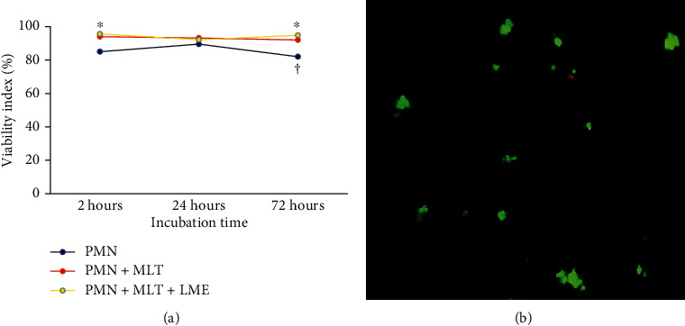
Time-dependent correlation of viability of PMN cells stimulated with melatonin (a). F = 9.26; p = 0.0018 (time of incubation), F = 14.4991; p = 0.0003 (treatment). The results represent the median of the standard deviation. PMN: polymorphonuclear cells; MLT: melatonin; LME: liquid microemulsion. The polymorphonuclear phagocytes were incubated with melatonin after 72 hours of incubation (a). Orange-stained cells (dead) and green-stained cells (alive). Experiments were repeated five times, and the results were comparable. p < 0.05. ∗Differences between PMN with treated PMN (MLT or MLT + LME), considering the same incubation times. †Differences between times of incubation, considering the same treatment.